
Blood In Urine (Haematuria)
Blood in the urine is known medically as haematuria. Haematuria is most commonly revealed during routine screening using a simple dipstick test. Haematuria can also be diagnosed when urine is examined under the microscope (microscopic haematuria) most commonly on a sample sent for urine culture for infection. Sometimes haematuria is visible, when the urine becomes red or brown in colour (macroscopic haematuria). Blood should not normally appear in the urine. Blood in the urine is a ‘red flag’ symptom and should always be investigated by an Urologist to exclude serious disease.Blood can come from any part of the urinary tract, from the kidneys, ureters, bladder, prostate gland or the urethra.
Incidence
Microscopic haematuria is found regularly on routine urinalysis. It is rare that patients under the age of 40 years with microscopic haematuria have a serious problem. In all other cases the presence of haematuria should be investigated. In many people, no specific cause is found. Cancer of the kidney or bladder is found in up to 5% of people with microscopic haematuria and up to 40% of those with visible haematuria.- loin pain
- 1-5% of patients with microscopic haematuria will have a malignancy
- 35% of patients with macroscopic haematuria will have an underlying tumour
Assessment (Haematuria Screen)
Haematuria always requires investigation by a specialist urologist. National guidelines mandate that standard sets of investigations are performed. These are listed below:- A physical examination is required including a prostate examination for men and gynaecological organs in the female
- Urine must be sent for
- Microscopy and culture (to exclude infection)
 Agar plate showing E.coli bacteria
 - Urine cytology (abnormal cells in the urine)
- It is important to note that this test only picks up cancer cells in 50% of patients with cancer
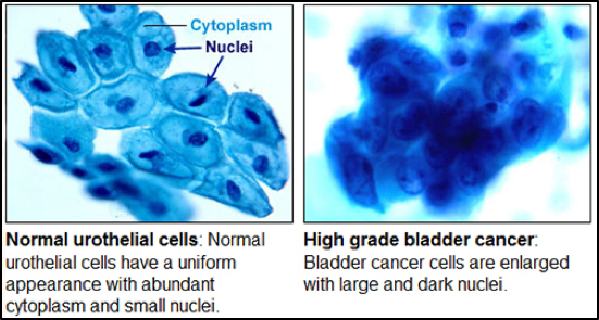 Image from John Hopkins University Pathology
- Microscopy and culture (to exclude infection)
- Imaging of the kidneysOur expert uro-radiologists specialise in imaging of the urinary tract and pelvis and work closely with their urology colleagues to provide the very best in diagnosis.
- Microscopic haematuria is usually investigated by Ultrasound
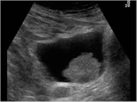 Ultrasound image showing bladder in black with bladder cancer growing inwards from bladder wall
 - Macroscopic haematuria is usually investigated by CT scanning
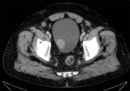 CT image showing bladder with bladder cancer growing inwards from bladder wall
- Microscopic haematuria is usually investigated by Ultrasound
- Inspection of the bladder (Cystoscopy). A fine fibre-optic camera is passed up the urethra (water-pipe) to view the lining of the bladder. The camera is passed under a short local anaesthetic and is not painful
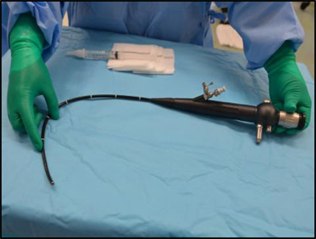 Flexible cystoscope, which is passed via the urethra under local anaesthetic to see inside the bladder. More information on the cystoscopy procedure can be found via the BAUS leaflet clicking here.